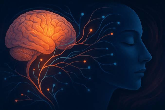
Scientists analyzed more than 6.3 million brain cells from 1,494 donors to map gene activity in the human prefrontal cortex across aging and disease.
A molecular map of the human brain at unprecedented scale
Scientists have produced one of the most detailed maps yet of gene activity in the human brain, analyzing more than 6.3 million cells from the brains of 1,494 people.
The work is part of a coordinated set of studies examining the prefrontal cortex, a region involved in planning, decision-making, emotional regulation and behavioral control.
The research was published on September 23 in a collection of papers across the Nature family of journals.
The significance is not simply the number of cells.
It is the combination of:
age + cell type + gene activity + disease + changes across the lifespan.

What does “gene activity” actually mean?
The researchers did not simply read people’s DNA.
They studied gene expression — which genes are active in different cells and how strongly they are expressed.
A neuron does not use the same genetic program as an immune cell.
Likewise, a brain cell in childhood does not necessarily have the same molecular profile as the same cell type in old age.
The researchers used single-cell and single-nucleus RNA sequencing to examine these differences at very high resolution.
One of the atlases contains 5.6 million nuclei from 1,384 donors of diverse ancestries.
From infancy to age 108
The project includes brain samples from people across a remarkably broad age range, from infancy to a person who was 108 years old.
That allowed researchers to study not only disease but also normal development and aging.
One major study analyzed 284 neurotypical donors ranging from infancy to age 97.
The researchers identified three broad phases of molecular change in the prefrontal cortex:
rapid early-life remodeling → relative stability → renewed remodeling in later life.
This does not mean the brain stops changing during midlife.
It means the dominant molecular patterns of change differ across stages of life.
Why the prefrontal cortex?
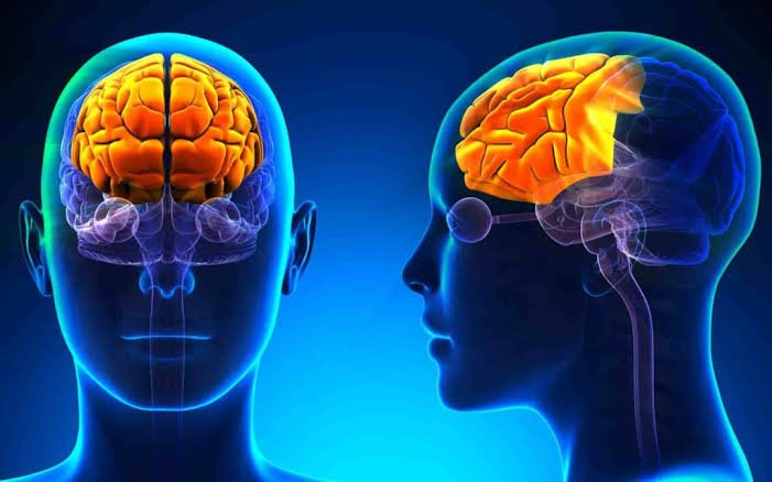
The prefrontal cortex is one of the brain’s most important regions for higher-order functions.
It contributes to:
- planning;
- working memory;
- decision-making;
- attention;
- behavioral control;
- emotional regulation.
It is also particularly vulnerable to age-related changes and is implicated in multiple neuropsychiatric and neurodegenerative disorders.

Alzheimer’s, Parkinson’s and schizophrenia in the same atlas
The project did not analyze only healthy or neurotypical brains.
The donor material included people diagnosed with:
- Alzheimer’s disease;
- Parkinson’s disease;
- Lewy body dementia;
- vascular dementia;
- tauopathy;
- frontotemporal dementia;
- schizophrenia;
- bipolar disorder.
That creates an opportunity to compare molecular changes across diseases and identify biological processes that are shared or disease-specific.
The NIH describes the effort as the largest comparative investigation so far of human brain disorders at single-cell resolution.
The brain’s molecular clock also changes
One of the studies found changes in circadian gene-expression patterns.
With aging, researchers observed reduced synchronization of neuronal clock-related gene expression and the emergence of stress-associated rhythms in glial cells.
Circadian rhythms influence many biological processes, including:
- sleep;
- metabolism;
- hormones;
- body temperature;
- nervous-system function.
But this does not mean the researchers discovered the cause of brain aging.
They identified molecular patterns that require further investigation.
More than 14,000 genes mapped
One of the atlases identified genetic regulation involving 14,258 genes.
Researchers also identified hundreds of cell-type-specific regulatory effects and more than 2,000 genes whose regulatory effects varied across developmental trajectories.
This is one of the major advantages of single-cell analysis.
When an entire piece of tissue is analyzed as one sample, molecular signals from different cell types can be mixed together.
Single-cell methods allow scientists to ask:
“What exactly is happening in this particular cell type?”

From molecular data to precision medicine
The long-term goal is not simply to create an enormous database.
The maps can help researchers identify biological mechanisms associated with disease and potentially discover new therapeutic targets.
But there is an important distinction:
A gene associated with a disease is not automatically the cause of that disease.
A molecular association may be:
- causal;
- consequential;
- contributory;
- or simply correlated with another biological process.
That is why the atlas should be viewed as a research foundation, not as a new treatment.
The NIH says the project is intended to support research into prevention, detection and more personalized treatment of brain disorders.
Why AI matters here
There is also a direct connection to technology.
Datasets containing millions of cells and thousands of molecular features are extraordinarily complex.
That is where:
bioinformatics + machine learning + statistical analysis + single-cell genomics
become increasingly important.
These tools can help identify patterns that would be extremely difficult to find manually.
That does not mean AI “discovered the brain atlas.”
Scientists generated the biological data and used computational methods to analyze and interpret it.
A “Google Maps” for the brain?
The analogy is imperfect, but useful.
A road map tells you where things are.
A molecular atlas tells you what is happening inside different cell types and how those processes change with age and disease.
The new atlas does not cover the entire brain.
It focuses on an important region — the prefrontal cortex — and much of the work relies on donated post-mortem brain tissue.
So this is not yet a real-time map of a living human brain.
That limitation matters.
What comes next?
The next step is to connect these findings with other regions of the brain.
The researchers themselves emphasize that the prefrontal cortex is only part of the picture. Understanding neurological disease fully will require data from additional brain regions and better connections between molecular changes and brain function.
The likely direction is:
larger atlases → more brain regions → more donors → better disease models → new therapeutic targets.
What readers should remember
Fact: Researchers analyzed more than 6.3 million brain cells/nuclei from 1,494 donors.
Fact: The project focuses on the prefrontal cortex, a region involved in higher cognitive functions.
Fact: Donors included people with Alzheimer’s, Parkinson’s, different forms of dementia, schizophrenia and bipolar disorder, alongside neurotypical controls.
Fact: One atlas includes 5.6 million nuclei from 1,384 donors and identifies genetic regulation involving 14,258 genes.
Interpretation: The dataset could help researchers identify molecular mechanisms associated with aging and brain disease.
Important: This is not a new cure for Alzheimer’s, Parkinson’s or schizophrenia. It is a large research foundation for future work.
Scientists have not yet created a complete map of the human mind.
But they have created something more concrete:
one of the most detailed molecular maps ever produced for a major region of the human brain.
Millions of cells, thousands of genes and people across the lifespan can now be studied within the same framework.
That gives neuroscience a more precise way to ask not only where the brain changes, but which cells and molecular processes are changing.
And that could define the next phase of brain research:
moving from “what disorder does the brain have?” toward “what exactly is happening in this cell type — and how can we change it?”